Microscopy

460 readers
1 users here now

Anything related to things that are too small to see them with the eye, and the tools used to observe them.

This space is quite general in scope - microscopes, microbiology, small component electronics, questions about buying optical components, etc.

founded 3 years ago
MODERATORS
26
34
submitted 9 months ago* (last edited 9 months ago) by Sal@mander.xyz to c/microscopy@mander.xyz


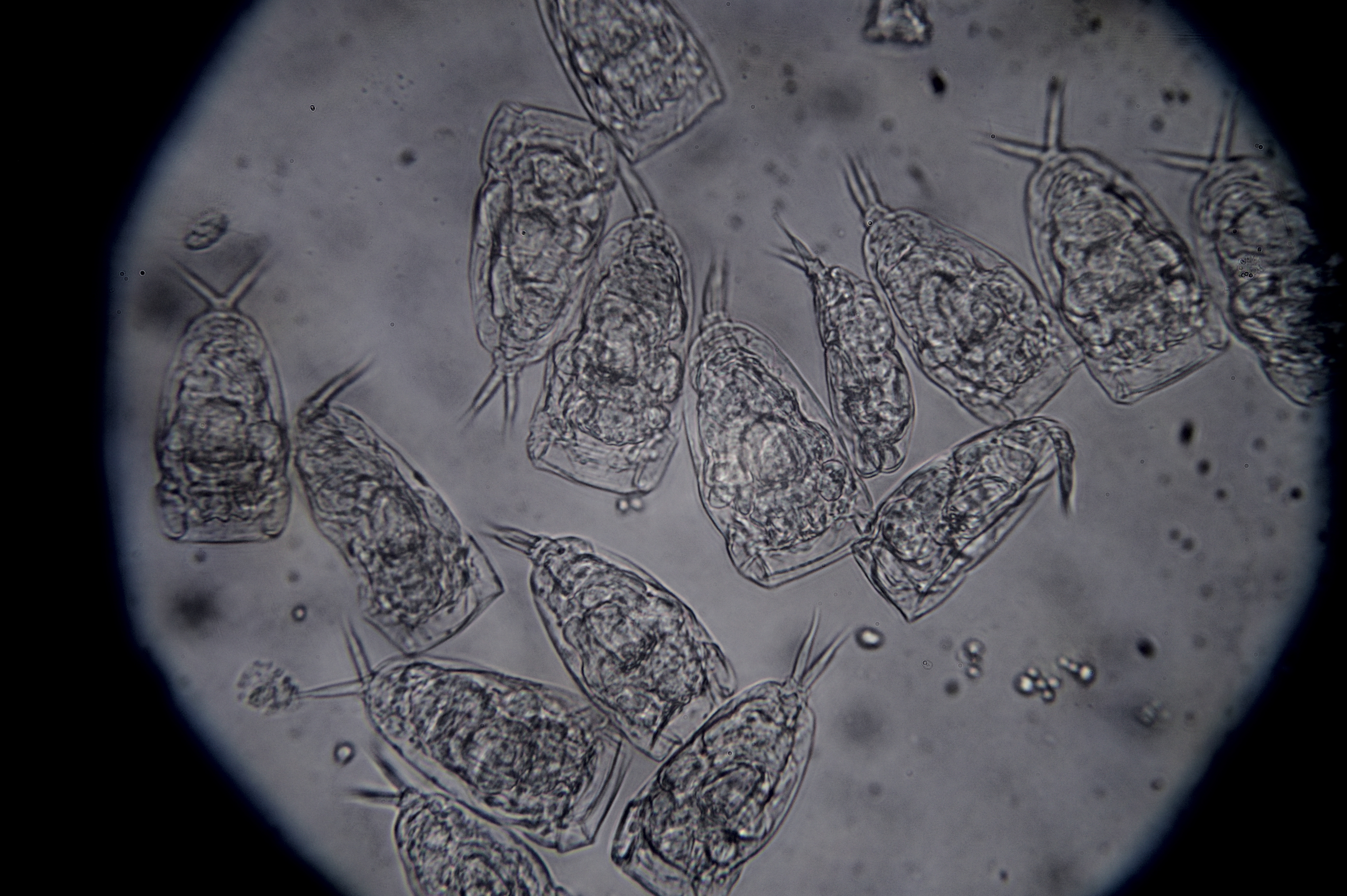
I prepared a 1:200 dilution of red blood cells using a ~1% NaCl solution. The imaged region contains 4 nano liters of the diluted sample. This image was taken using a 40x objective.

A count is performed by counting the number of red blood cells in a few of these sections, averaging the result, and then converting back to red blood cells per microliter by multiplying times 200 (dilution) and dividing by 0.004 (sampled volume in micoliters).

For this particular sample I estimated 3.8 million red blood cells per micro liter of blood.

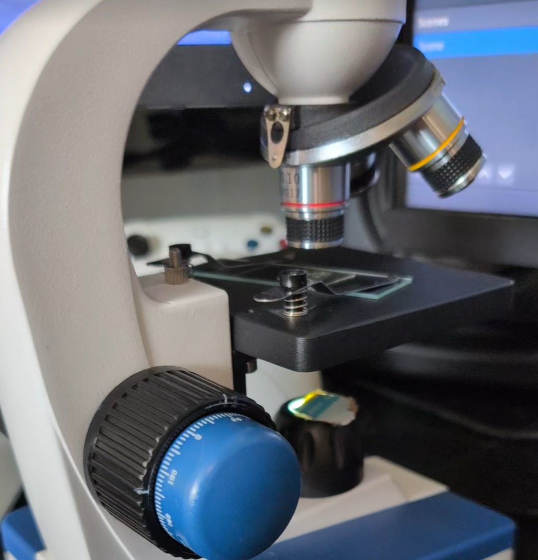
I tested a few different types of hemocytometer/Neubauer chambers from China and I can recommend this specific one:

There are some even cheaper alternatives but the lines are very difficult to see.

27
13
Epithelial cells (mander.xyz)
submitted 9 months ago* (last edited 9 months ago) by Sal@mander.xyz to c/microscopy@mander.xyz


I followed the Gram Staining tutorial from this video to prepare a sample of my cheek cells: https://www.youtube.com/watch?v=lMoT-FmhS6A

For preparing the staining solutions I purchased crystal violet, ethanol, potassium iodide, iodine, and an already prepared safranin solution from laboratorium discounter.

The slight 3D effect is achieved by displacing the filter holder to block the light coming from one direction and achieve oblique illumination to cast a shadow (https://www.youtube.com/watch?v=9btIpf5mjyA).

The image is post-processed using Rawtherapee to increase the contrast.

Here is another photo without using the oblique illumination trick, also post-processed with rawtherapee:

28


In trying to isolate Trebouxia from an Evernia lichen. I found that some of the cultures are contaminated by a what I think are rotifers. I am not sure of what kind of rotifer (or other organism) is the one pictured, so if anyone has some idea please let me know.

I also recorded a video of what I think are belloid rotifers feeding on the same lichen culture:

https://peertube.uno/w/uoSCNagVVmbuMcgXdVfPGR

I don't have much hope that the algae will survive this attack, but I might turn those jars into rotifer cultures.

29


I left a slide with some algae and rotifers sitting on the microscope. After it dried up I was able to see several of these flower-like shapes. Not a pattern that I had seen before, and I a don't know what about the drying process lead to this particular shapes forming.

30
 
 
31


I'm very happy with my fake guitar sound.

YouTube link: https://youtu.be/bZNL08qahOQ

32
 
 
33


I will be making sure they are the right objectives in every way before I purchase anything. I'd just like to know where I can start looking. I added a 60x a while back, it was really cheap but works surprisingly well considering I payed like 20$ CAD for it. Parfocality is just about the same as my stock objectives, just a slight turn of the fine focus. That said, I'd like to spend more and get a whole new set of higher quality objectives.

I'd love to hear feedback if someone here has switched out objectives on this particular model. I do not need to hear that my microscope sucks haha, I'm a hobbyist so its not a big concern of mine at this time.

34
 
 
35
 
 
36
 
 
37
 
 
38


I am now uploading to peertube too!

Speedy Biofilm https://peertube.wtf/videos/watch/39f8e00e-2313-4cc3-9e91-d1f4c7fc6a04

39
40
41
42
43
44
45


This one just shot out of me. Idk how it happened so fast. I've gotta slow down at some point. I was really excited to show y'all my first attempt at darkfield.

Here's some pics to show how I did it.

46
47
48
49
50
view more: ‹ prev next ›